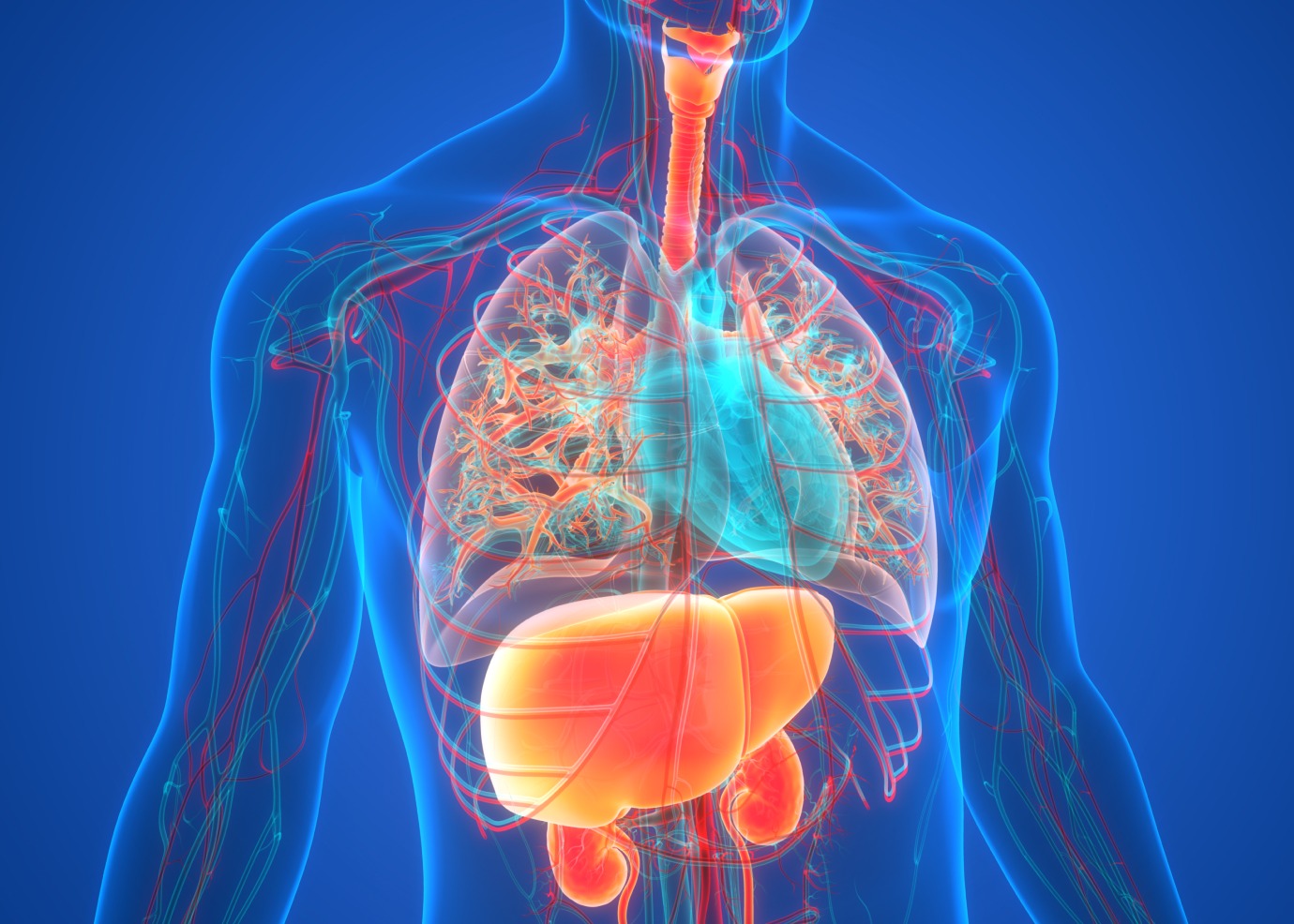
Why is our heart to the left and the liver to the right?
Heart to the left. Liver to the right. That’s where you’ll find these organs in a healthy human body. But surprisingly, in some people, the heart is on the right and the liver on the left. This normal or abnormal asymmetry can be traced back to your embryonic stage. In the early days of your development, a small fluid-filled cavity known as an embryonic node formed on your embryo. Inside, tiny micro-hairs known as cilia create a flow pattern that steers where organs grow in your body. However, the science behind this flow process has remained a mystery until now. Researchers from Eindhoven University of Technology (TU/e) and the University of Groningen have revealed key details of the process by building a world-first artificial embryonic node that uses synthetic magnetically controlled cilia to generate a flow pattern. They explored what happens in the node using advanced simulations. Their findings are published in the journal Science Advances.
FSE Science Newsroom | Text TU/e
On the outside, the human body is bilaterally symmetrical. A face with two eyes and two ears that mirror each other. Limbs that do the same. Inside the body is a different story. While paired organs, such as lungs and kidneys, are roughly bilaterally symmetrical, the same can’t be said about the other organs. We don’t have two hearts or two livers. What’s more, you’ll find these organs on the opposite sides of the body – the heart on the left, and the liver on the right. So, what is the origin of this organ asymmetry?

Embryonic origins
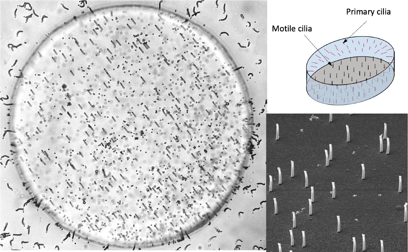
‘It can be traced all the way back to your first period as an embryo,’ says Jaap den Toonder, professor in the Department of Mechanical Engineering and chair of the Microsystems section at TU/e. ‘And it has to do with what happens in something called the embryonic node.’ This embryonic node is a small cavity filled with fluid (water, proteins, hormones, and other substances). The top is closed off by a membrane, while the bottom layer is lined with a few hundred tiny micro-hairs called cilia. The node is just a few hundred micrometers across.
‘The cilia in the embryonic node rotate in the same direction, making a tilted conical motion. This generates an anticlockwise fluid flow inside the node, and it’s this flow that is known to play a key role in the left-right symmetry,’ explains Den Toonder.
A critical question that remains unanswered is how exactly the flow instigates the left-right distribution of organs in the body. To solve this riddle, he led a project to design an artificial embryonic node - a world first. Developing the artificial embryonic node were Tanveer ul Islam, Yves Bellouard (associate professor at EPFL, Switzerland), and Den Toonder, while Ishu Aggarwal and Patrick Onck (both at the University of Groningen) led efforts to develop a model to simulate the embryonic node.
Delicate fabrication process
‘Fabrication of the artificial embryonic node involved two key micro-fabrication processes,’ says Tanveer ul Islam. ‘In the first process, we made the cilia from a unique magnetic polymer material that was developed at the TU/e. The second process involved making the node itself.’
While the artificial embryonic nodes are about five times larger than real embryonic nodes (about 500 micrometers across), they are still at a length scale comparable to real embryonic nodes. ‘The reason for this size is that light microscopy could not accurately visualize all the motion of all cilia and the fluid if the node were smaller,’ adds Tanveer ul Islam, who did much of the work.
Next, Tanveer ul Islam and his colleagues designed a way to control the magnetic cilia while observing the flow pattern created inside the node at the same time. ‘We developed a new magnetic actuation method to manipulate the motion of the cilia, while allowing a high-resolution recording of the fluid flow using fluorescent tracer particles. Without this visualization approach, we would not have been able to decipher how the embryonic node works,’ he explains. Visualizations revealed how the cilia cause very specific flow patterns.

Two mechanisms working together
Toonder: ‘Several hypotheses had been proposed for the asymmetric development. One is that the cilia at the bottom generate the flow, and then the other, non-motile, cilia known as primary cilia and located at the edges inside the node, detect the magnitude of the flow by deforming. This, in turn, triggers a genetic program steering the asymmetric organ development. Another hypothesis is that this genetic program is instead initiated by the nodal flow, inducing an asymmetric distribution of signaling molecules called morphogens.’
To test these hypotheses in the artificial embryonic node, the numerical model of Ishu Aggarwal and Patrick Onck at the University of Groningen was instrumental. ‘Jaap and I have been working together for over 20 years on experiments and simulations related to magnetic artificial cilia,’ says Onck. The combination of the experiments and the simulation revealed that both mechanisms, the primary cilia bending and the morphogen distribution, work together, suggesting a synergy between the two sensing mechanisms to drive the left-right asymmetry of organ development.

Combined approaches were essential
To date, no groups have been able to simulate the complete embryonic node as current models require vast computational power and resources. ‘To simulate the fluid flow in the embryonic node, the cilia were modeled as rigid rods that exactly follow the time-varying magnetic field from the experiments. By placing the cilia at the measured positions, we were able to accurately simulate the cilia-induced flow pattern from the experiments.’
The researchers at Groningen used their own in-house algorithm to simplify the simulation of fluid flow in the embryonic node. ‘We created a set of equations to approximate the fluid and remain solvable in a feasible amount of computational time. Despite this, the simulations still took months of computer time on our high-performance supercomputing facilities of the University of Groningen,’ adds Onck.
‘The main thing to note is that we captured the induced flow together with the convection and diffusion of morphogens within the fluid in the embryonic node, as well as the small-scale deformation of the primary cilia. In combination with the experiments, these combined approaches were essential to show what breaks the left-right symmetry, namely the combination of the two synergetic mechanisms.’
Reference: Tanveer ul Islam, Ishu Aggarwal, Yves Bellouard, Patrick R. Onck, and Jaap M. J. den Toonder: Artificial embryonic node elucidates the role of flow in left-right symmetry breaking in vertebrates. Science Advances 25 March 2026