
Betere resolutie voor bepaling weefselstijfheid via MRI dankzij wiskunde
Al eeuwen bepalen artsen de stijfheid van weefsels, bijvoorbeeld bij onderzoek naar borst- of prostaatkanker, met de hand. Maar alleen moderne beeldvormende technieken, zoals magnetische resonantie elastografie (MRE) zijn geschikt om dit op een niet-invasieve manier te doen in bijvoorbeeld lever of hersenen. RUG-wetenschappers hebben, samen met collega’s uit Chili en Duitsland, een wiskundige theorie en een algoritme ontwikkeld waarmee ze enkele problemen die MRE met zich meebrengt op kunnen lossen. Een test met MRI data verzameld bij mensen laat zien dat deze aanpak veelbelovend is, en hoge resolutie beelden van de stijfheid van hersenweefsel produceert. Dit onderzoek is gepubliceerd in het tijdschrift Medical Image Analysis.
MRE is een veelbelovende techniek om op een niet-invasieve manier kwantitatieve informatie te verzamelen over de elasticiteit van zacht weefsel. De principes van deze methode zijn in 1995 beschreven door wetenschappers van de Mayo Clinic (VS), en sindsdien is deze nuttig gebleken bij het volgen van het verloop van verschillende ziekten, zoals leverfibrose en neurologische aandoeningen. Met een mechanisch apparaat stuurt de onderzoeker een trillingsgolf door het weefsel, terwijl er beelden worden gemaakt in een MRI scanner. Met behulp van natuurkundige en wiskundige principes die golftransport beschrijven is vervolgens de stijfheid van het weefsel te bepalen.
Ruis
‘Het is uitdrukkelijk niet zo dat de MRI scanner een direct beeld maakt van de golf’, zegt Cristóbal Bertoglio, universitair docent in Wiskunde, Computerwetenschap en Kunstmatige Intelligentie bij het Bernoulli Instituut van de RUG. ‘De scanner produceert en meet een magnetisch veld in het weefsel, dat wordt weergegeven door een vector met een tollende beweging, precessie genaamd. De snelheid van deze precessie neemt toe met de amplitude van de golf en de kracht van het magnetisch veld dat de MRI scanner produceert’, zo legt hij uit. ‘Een complicatie is dat je niet kunt zien of de hoek hoort bij de eerste, tweede of volgende rotatie omdat je niet weet wat de amplitude oorspronkelijk was.’
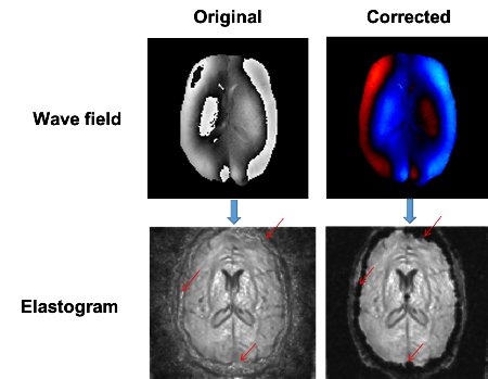
Een mogelijke oplossing is om een erg laag magnetisch veld toe te passen, wat de hoek verlaagt en er voor zorgt dat de meting in de eerst rotatie valt. Maar dit versterkt de ruis in het beeld dat van de golf wordt gemaakt, zodat de kwaliteit van de stijfheidsmeting sterk afneemt. Dit probleem is weer aangepakt door twee aparte beelden te maken bij verschillende magnetische veldsterkten, die vervolgens worden gecombineerd. Maar er bestaan geen theoretische modellen die beschrijven hoe dit het beste kan gebeuren. ‘Daarom gebeurt het vooral door maar verschillende aanpakken te proberen, maar dat levert fouten op in de afbeelding van de stijfheid’, legt Bertoglio uit.
Meer detail
Daarom ontwikkelde Bertoglio, samen met Helge Herthum van de MRE-groep in het Charité Universiteitsziekenhuis van Berlijn en met voormalig promovendus Hugo Carrillo een algemene theorie die beschrijft hoe verschillende metingen zijn te combineren. ‘We begonnen met een algemene MRI theorie en leidden hieruit een deeltheorie af die de combinaties beschrijft. Op die manier produceerden we een theoretisch model dat werkt.’ Dit model is getest op echte MRE data van hersenweefsel, waarin metingen zijn gecombineerd, gedaan bij verschillende sterkten van het magnetisch veld. Het resultaat was een afbeelding met weinig ruis en geen artefacten. ‘We kunnen nu betere beelden van de elasticiteit maken zodat we de stijfheid van het weefsel kunnen meten in meer detail.’
Veelbelovend
Bertoglio gaat nu samen met biologen, radiologen en neurochirurgen van het UMCG aan de slag om met deze nieuwe techniek de stijfheid van hersentumoren te bepalen. ‘Tumoren kunnen hard of zacht zijn’, legt hij uit. ‘En daarvoor zijn verschillende chirurgische benaderingen nodig. Het is daarom belangrijk om voor de operatie via een niet-invasieve manier de stijfheid van de tumor te bepalen.’ Een andere toepassing is het afleveren van geneesmiddelen met moleculaire transportsystemen. ‘Het type systeem dat je kiest hangt mede af van de stijfheid van het weefsel waar je de medicijnen in wilt afleveren.’
Bertoglio begon te werken aan een wiskundige model voor MRI scans toen hij nog verbonden was aan het Centre for Mathematical Modelling in Santiago, Chili. Daar werkte hij samen met de hoogleraren Axel Osses en Sergio Uribe. ‘Dit werk was gericht op het combineren van twee metingen van de stroomsnelheid van bloed.’ Vervolgens stapte hij over naar de RUG, en kwam in contact met prof. Ingolf Sack van de Charité groep. ‘We zagen hoe nodig en hoe veelbelovend MR elastografie was. Verder was het mooi dat we Helge al vanaf het begin aan boord hadden. Dit leverde een geweldig interdisciplinair en internationaal team op, dat deze resultaten mogelijk heeft gemaakt.’
Referentie: Helge Herthum, Hugo Carrillo, Axel Osses, Sergio Uribe, Ingolf Sack and CristóbalBertoglio: Multiple motion encoding in phase-contrast MRI: A general theory and application to elastography imaging. Medical Image Analysis, online 14 maart 2022.
Meer nieuws
-
-
02 juni 2026
Minuscuul reukspoortje ontrafeld
-
01 juni 2026
Materiaal met eigenschappen bepaald door structuur