
Mathematics increases the resolution of MRI measurements of tissue stiffness
Manual palpation has been used diagnostically for centuries to assess tissue stiffness, with breast and prostate cancer screenings being well-known contemporary examples. However, only advanced imaging techniques, such as magnetic resonance elastography (MRE), enable non-invasive mapping of liver and brain tissue mechanics. Mathematicians at the University of Groningen in the Netherlands, in collaboration with scientists from Chile and Germany, have developed a mathematical theory and algorithm that overcome some of the key challenges associated with this technique. The resulting human MRI data are promising, as evidenced by more detailed, high-resolution reconstructed images of stiffness in brain tissue published in Medical Image Analysis.
MRE is an emerging technique used for non-invasive and quantitative imaging of the viscoelastic properties of soft tissue. Since scientists at the Mayo Clinic first formulated the principles of MRE in 1995, the technique has advanced and proven effective for monitoring the progression of various diseases, including liver fibrosis and neurological degradation. A vibration wave is transmitted through the targeted tissue using a mechanical device and imaged with an MRI scanner. Stiffness of the target tissue is subsequently inferred using physical and mathematical laws of wave propagation.
Noise
‘However, it is important to note that the MRI scanner does not directly make an image of the wave’, says Bertoglio, assistant professor in mathematics at the Bernoulli Institute for Mathematics, Computer Science and Artificial Intelligence, University of Groningen. ‘The scanner actually produces and measures a magnetic field in the tissue, represented by a precessing vector. But the precession speed increases with both the amplitude of the wave and the strength of the magnetic field applied by the MRI’, he explains. ‘A major challenge is that you can’t be sure whether the vector’s angle corresponds to the first, second, or more laps, as you do not know, a priori, the actual amplitude of the wave’.
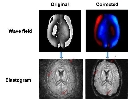
A possible solution could be to apply a very low magnetic field to reduce the angle, thereby ensuring that it corresponds to the first lap. However, the noise in the image of the wave then becomes amplified, leading to a considerable decline in the quality of the stiffness maps. This problem was tackled by measuring two separate images at different magnetic strengths and then combining them. However, no theoretical model existed for combining these two measurements. ‘Therefore, the combination was usually done by trial and error, which led to the generation of artefacts in the resulting elasticity maps’, Bertoglio explains.
Details
In collaboration with Helge Herthum, who is a member of the MR Elastography Group at Charité Universitätsmedizin in Berlin and Hugo Carrillo, his former PhD student, Bertoglio developed a general theory for establishing combinations of different sets of measurements. ‘We started from general MRI theory and derived a sub-theory, which describes the combinations. This way, we created a theoretical model that made sense.' This theoretical model was then tested on a set of actual MRE brain data, in which measurements were combined using several magnetic fields of varying strengths. The result was a low-noise map without artefacts. ‘We can now generate better elasticity maps to measure the stiffness of tissues, which allow us to see more details.'
Potential
Bertoglio plans to work with biologists, radiologists and neurosurgeons at the University Medical Center Groningen, using the new technique to assess brain tumours. He explains: ‘Tumours can be hard or soft, and require different surgical approaches to remove them. It is therefore very useful to use a non-invasive method to determine the stiffness of the tumour before an operation’. Drug delivery using nano-carriers could be another potential application: ‘The type of carrier that should be used may also depend on the stiffness of the tissue’.
Bertoglio began developing mathematical models of MRI scans while at the Centre for Mathematical Modelling in Santiago, Chile, where he worked in close collaboration with Professors Axel Osses and Sergio Uribe. ‘This work focused on the combination of two measurements in blood flow imaging’. He later moved to Groningen and established contact with Professor Ingolf Sack from the Charité group. ‘We recognised the great need and potential of this technique in MR elastography, and we were very lucky to have Helge on board from the beginning. We had a great interdisciplinary and international team of collaborators, which made these results possible.’
Reference: Helge Herthum, Hugo Carrillo, Axel Osses, Sergio Uribe, Ingolf Sack and CristóbalBertoglio: Multiple motion encoding in phase-contrast MRI: A general theory and application to elastography imaging. Medical Image Analysis, online 14 March 2022.
| Last modified: | 07 February 2025 12.32 p.m. |
More news
-
06 May 2025
Overcoming grid congestion: ‘Making better use of what we already have’
Grid congestion poses a major problem. There is little to no capacity to connect new households and businesses to the power grid and it risks halting the energy transition. Michele Cucuzzella, Associate Professor of Energy Systems & Nonlinear...
-
29 April 2025
Impact | Rubber recycling
In the coming weeks the nominees for the Ben Feringa Impact Award 2025 will introduce themselves and their impactful research or project. This week: Francesco Picchioni on his innovative way to recycle rubber.
-
29 April 2025
Impact | Improving Human-AI Decision-Making in healthcare
In the coming weeks the nominees for the Ben Feringa Impact Award 2025 will introduce themselves and their impactful research or project. This week: Andra Cristiana Minculescu on her research project on Human-AI Decision-Making in healthcare.