Optisch pincet onthult virus zelf-assemblage proces
Terwijl we inmiddels heel goed weten hoe virussen opgebouwd zijn, blijft het onduidelijk hoe virussen gevormd worden. Fysici hebben nu een tipje van de sluier opgelicht, door het virale assemblage proces “live” te volgen. Deze tour-de-force was mogelijk door één enkel DNA molecuul op te hangen, met behulp van een zogenaamd optisch pincet, en te kijken hoe de viruseiwitten stuk voor stuk aan het DNA hechtten. Hierdoor konden niet alleen de eerste stappen van zelf-assemblage worden blootgelegd, maar ook het hierop volgende groeiproces.
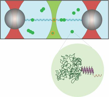
De onderzoekers uit Mexico, Eindhoven, Wageningen, Amsterdam en Groningen gebruikten het optisch pincet, een techniek waarvoor in 2018 de nobelprijs in de natuurkunde werd uitgereikt, om een DNA molecuul tussen twee bolletjes met behulp van een laserstraal te vangen. Door daarna lichtgevende eiwitten van een kunstmatig virus toe te voegen, kon het aanhechtten aan en inpakken van het DNA gevolgd worden. De figuur verduidelijkt het proces. Zernike onderzoeker en laatste auteur, Wouter Roos: ,,Het mooie van deze techniek is dat je niet alleen kunt zien waar de eiwitten vast gaan zitten, maar je ook de kracht kunt meten die op de bolletjes wordt uitgeoefend. Hierdoor kan je heel precies bepalen hoe het DNA door het virus wordt ingepakt.” De metingen werden ondersteund door andere geavanceerde technieken, nl. Acoustic force spectroscopy (AFS) en Atomic force microscopy (AFM). AFS is een techniek waarbij de bolletjes door geluidsgolven, ipv door licht, worden bewogen en AFM is een tastmicroscoop. Door de data van deze drie technieken te combineren, konden de onderzoekers bepalen dat het inpakken met hele regelmatige stappen gebeurd. Nog niet eerder was deze regelmaat aangetoond.
De nieuwe inzichten op het gebied van virusassemblage brengen niet alleen het fundamenteel onderzoek verder. Door beter te begrijpen hoe virussen in elkaar worden gezet, wordt het ook makkelijker om gericht naar anti-virale middelen te zoeken die specifiek virus assemblage blokkeren.
Meer informatie:
Margherita Marchetti, Douwe Kamsma, Ernesto Cazares Vargas, Armando Hernandez García,
Paul van der Schoot, Renko de Vries, Gijs J. L. Wuite, and Wouter H. Roos
Real-Time Assembly of Viruslike Nucleocapsids Elucidated at the Single-Particle Level
Nano Letters 2019, Vol. 19, 5746−5753