Skip to ContentSkip to Navigation
Research Zernike (ZIAM) News

Looking Inside Organic Solar Cells

08 November 2016
Conceptual representation of the all-optical method to retrieve morphology. A laser pulse (green beam) creates a quasi-particle, the exciton (blue-red circle). The exciton diffuses (purple arrow) within the acceptor domain (yellow hills) and dissociate into the electron (blue circle) and hole (red circle) at the interface with the donor material (red valleys). The concentration of the holes is probed by the second delayed pulse (brown beam). The acceptor domain size is retrieved from the delay between exciton creation and dissociation.

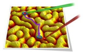
Organic solar cells form an emerging area of photovoltaics, which promises to substitute traditional silicon solar modules for niche applications. To convert the sun energy into electricity in such a device, a bulk heterojunction is used, which is a phase-separated mixture of donor- and acceptor-type organic semiconductors. Achieving the highest power conversion efficiencies requires thorough control of the nanoscale structure of the bulk heterojunction, the so-called morphology. As the typical phase separation of the two materials occurs in 10 nm region, the morphology is extremely hard to study by conventional microscopic techniques.

The Optical Condensed Matter Physics and Polymer Science groups at the Zernike Institute for Advanced Materials have recently developed an all-optical method to determine key information on the morphology of the bulk heterojunction. Using a combination of ultrafast spectroscopy and kinetic Monte-Carlo simulations, they captured morphology features with nanometer spatial resolution. Independent verification of morphology reconstruction by microscopic and surface-sensitive X-ray scattering techniques demonstrated the validity of the all-optical approach. As the proposed technique is non-invasive, it can be used for on-the-fly characterization of fully functional devices thereby opening exciting perspectives for developing high-efficient organic optoelectronics.

The results of this work are published in

Serbenta, A. et al. Bulk heterojunction morphology of polymer:fullerene blends revealed by ultrafast spectroscopy. Sci. Rep. 6, 36236; doi: 10.1038/srep36236 (2016)

www.nature.com/articles/srep36236

Last modified:08 November 2016 1.19 p.m.

More news

  • 16 April 2024

    UG signs Barcelona Declaration on Open Research Information

    In a significant stride toward advancing responsible research assessment and open science, the University of Groningen has officially signed the Barcelona Declaration on Open Research Information.

  • 02 April 2024

    Flying on wood dust

    Every two weeks, UG Makers puts the spotlight on a researcher who has created something tangible, ranging from homemade measuring equipment for academic research to small or larger products that can change our daily lives. That is how UG...

  • 18 March 2024

    VentureLab North helps researchers to develop succesful startups

    It has happened to many researchers. While working, you suddenly ask yourself: would this not be incredibly useful for people outside of my own research discipline? There are many ways to share the results of your research. For example, think of a...