MicroPET
FACILITY FOR SMALL ANIMAL IMAGING
(microPET / microSPECT / microCT)
Positron emission tomography or PET is a unique imaging modality providing quantitative data on cell-cell communication, tissue function and metabolism. MicroPET cameras with a small field of view and superior spatial resolution (1-1.5 mm) allow application of the PET technique to studies in small animals like rodents.
Even higher spatial resolution (up to 0.35 mm) is possible with single photon emission tomography (SPECT) using a multi-pinhole camera such as the U-SPECT II. The SPECT technique allows study of the in vivo behavior of radiolabeled peptides and antibodies, e.g. in tumor-bearing animals. Anatomical information (acquired with a microCT) can be combined with metabolic information (acquired with microPET or microSPECT) in order to facilitate data interpretation.
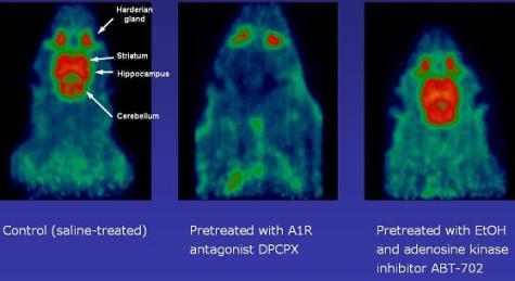
Radiopharmaceuticals for assessment of blood flow, metabolism, enzymes, receptors, transporters, angiogenesis, apoptosis, beta-amyloid plaques and neurofibrillary tangles make it possible to employ PET in basic neuroscience, oncology and cardiovascular research. In animal models of human disease, PET can be used for quantification of gene expression and to assess the pharmacokinetics and pharmacodynamics of drugs. Basic research on the in vivo trafficking of stem cells and of inflammatory cells may also benefit from animal PET technology. The Groningen Facility for Small Animal Imaging has been initiated with funding from NWO. It is supported by the Department of Nuclear Medicine and Molecular Imaging of the UMCG which has a large expertise in radiochemistry and pharmacokinetic modelling. Since the Facility has national importance it is accessible to researchers from industry and academia throughout the Netherlands
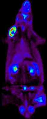
![Left: microPET image, right: microCT image, middle: fused microPET/CT images of a mouse bearing CT26 (b) and CT26mßGUS tumors (a). The microPET image was made with a novel ß-glucuronidase tracer, [18F]-FEAnGA](https://www.rug.nl/umcg/faculteit/disciplinegroepen/straling/ngmb/image/MicroPET-CT-Image.jpg)