The microPET, microSPECT and microCT scanners
An area for animal experiments has been created in the basement of the Department of Nuclear Medicine & Molecular Imaging (NMMI) of the UMCG.
The imaging devices available are a microPET, microCT and a microSPECT.
The same rodent bed fits into all scanners, thus metabolic and anatomic information (PET or SPECT and CT images) can be easily combined.
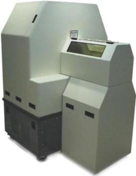
Parameters of the Siemens microPET Focus 220 scanner for small animals:
|
Detector diameter (cm) |
26 |
|
Effective opening (bore) (cm) |
22 (26 cm with shields removed) |
|
Transaxial Field of View (FOV in cm) |
19 |
|
Axial Field of View (cm) |
7.6 |
|
Number of detector blocks |
168 |
|
Total number of LSO elements |
24,192 |
|
Pixel element size(mm3) |
1.5 x 1.5 x 10 |
|
Absolute system sensitivity (%) |
4.0 |
|
Resolution at center of FOV (mm) |
< 1.3 |
|
Average energy resolution (%) |
< 18 |
|
Volumetric resolution (center FOV) (µl) |
< 2.5 |
|
Volumetric resolution (central 8 cm) (µl) |
< 9 |
|
Variable timing window: |
2, 6, 10, 14 ns |
|
Variable energy window: |
0 – 814 keV |
|
Set of 2D and 3D reconstruction algorithms |
|
|
Computer controlled bed |
|
|
Attenuation correction |
|
|
Deadtime correction |
|
|
Scatter correction |
|
|
Decay correction for the most applied radionuclides |
|
|
Dynamic framing |
(from 1 ms to static) |
|
Respiratory and cardiac gating |
|
|
Interchangeable beds for mice and rats |
For data acquisition, sinogram and image analysis, various software packages are available.
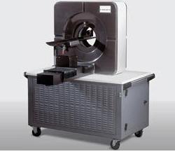
A Siemens Imtek MicroCAT II (small animal CT scanner) is placed in the same room as the microPET Focus 220. The CT is installed in a fully shielded cabinet x-ray system as defined by FDA Standard 21, CFR 1020.40. Parameters of that system are:
|
X-ray source |
SourceRay SB-80-50 |
|
Maximum Potential |
80 kVp |
|
Maximum current |
0.5 mA |
|
Anode material |
Tungsten |
|
Focal spot size |
~ 40 µm |
|
Beam filtration |
0.25 – 2 mm Aluminium |
|
Duty cycle |
100 % |
The gantry port is configured with lasers to draw calibrated x,y,z fiducial markers on the animal for easy positioning. X-ray dose depends on many factors but for a typical whole animal scan of 360 steps (rat) the delivered dose is < 10 cGy.
The detector is a CCD camera with 2048 x 3072 pixels and 5 frame per second read out with 2x2 binning. Dynamic range: 69 dB with 1 x 1 binning and 72 dB with 2 x 2 and 4 x 4 binning.
|
Standard position |
57 x 85 mm2 FOV |
|
37 µm resolution |
|
|
High resolution position |
31 x 47 mm2 FOV |
|
27 µm resolution |
The microCAT reconstruction software package is based on a modified Feldkamp algorithm weighted for either 180-degree plus cone-beam or 360-degree studies. The microCAT system has been delivered with the Amira Advanced 3D Visualization and Volume Modelling package.
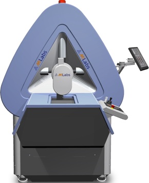
Micro-SPECT system based on a stationary detector set-up, resulting in:
· high mechanical stability
· low maintenance
· high up time
Quantitative image reconstruction software
Sub-minute temporal resolution
Energy range 20-400 keV
List mode acquisition
Reconstructed resolution down to:
· 0.6 mm (standard mouse collimator)
· 1 mm (rat collimator)