Electroshock treatment of depression generates new nerve cells
The treatment of depression with electroconvulsive therapy (ECT, previously often referred to as electroshock therapy) stimulates the generation of new nerve cells. The ECT causes a specific part of the brain, the dentate gyrus, part of the hippocampus, to expand. This is the only part of the adult human hippocampus that is capable of producing new nerve cells. Increase in this structure lead to a reduction in depressive complaints. This is demonstrated by research performed by brain scientists at the University Medical Center Groningen (UMCG) and University Medical Center Utrecht (UMCU), under the direction of University of Groningen Professor Iris Sommer. This research is the first to provide insight into the specific reasons why ECT is such an effective treatment for depression. The results were published today in the prominent scientific journal Molecular Psychiatry.
Electroconvulsive therapy (ECT) has been used by psychiatrists for more than 75 years. During this time, much has changed about the conditions under which the procedure is applied – today, patients are put to sleep, given muscle relaxants and supervised closely. The essential process, however, remains the same: inducing an epileptic stroke by sending an electric current through the brain. ECT has remained in use as a treatment because of its particular effectiveness in severe cases of depression. In the field of psychiatry, it is one of the most effective treatments available. It was already a well-known fact that the treatment works, but until now, it was not yet entirely clear as to why this is the case.
Research using a high-capacity MRI scanner
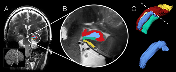
New research, performed by first author Jasper Nuninga and fellow scientists at the UMCG and UMCU, casts new light on the question of how ECT affects the brain. In the study, depression patients who receive ECT treatment underwent MRI scans both before and after their treatment sessions. A control group also underwent two scans. During these scans, the focus in was on the hippocampus, a small structure in the brain which is important for human thought and action. In many brain disorders, with Alzheimer’s disease the most prominent among them, the hippocampus is in a shrunken state. The same is true the case in depression, although this is much less pronounced than in case of Alzheimer’s.
ECT causes enlargement of one specific brain region
The hippocampus consists of different parts, each of which have a different function. Only one of these parts, the dentate gyrus, is capable of producing new nerve cells (this process is called neurogenesis). The study showed that the dentate gyrus was considerably enlarged after ECT treatment. This size difference before and after treatment offers an indication of the beneficial effect of ECT on depression, as a relationship was found between the increase of the dentate gyrus and the alleviation of depressive complaints: the greater the increase in size, the greater the treatment’s effectiveness. This offers a significant indication that the effectiveness of ECT can be explained by the fact that it stimulates the generation of new nerve cells. This is a very interesting finding, partly because it suggests the possibility that neurogenesis might also be stimulated by other means, which might lead to different forms of treatment for depression.
Source: press release UMCG
More news
-
26 May 2026
Impact |Safer PET/CT imaging for children
-
-