New microscope shows the atomic structure of materials in stunning detail
A new transmission electron microscope (TEM) allows University of Groningen scientists to study the structure of materials in unprecedented detail. One of its unique abilities is to produce images of both heavy and very light atoms simultaneously. The microscope will be primarily used by the Zernike Institute for Advanced Materials (ZIAM) and CogniGron research institutes, but biologists might also take advantage of this new piece of equipment.
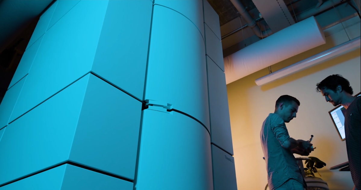
The four-metre-tall microscope has been installed in a climate-controlled room in the basement of the UG Physics and Chemistry building at the Zernike Campus. ‘This was the only room where it would fit,’ says Professor Bart Kooi, chair of the Nanostructured Materials and Interfaces research group and the person responsible for the running of the new microscope.
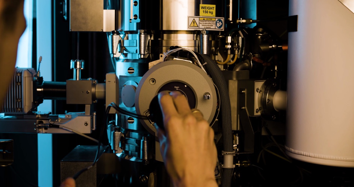
The machine itself is hidden behind white panels and remotely operated. ‘We have an operator room next door, but we can also access the machine from the other side of this building.’ After loading the samples in the microscope, the system should be left alone for a while. Studying matter at atomic level requires a very controlled environment. Electron microscopes have been around for decades. They use electron beams, rather than light, to create an image. The sample changes the trajectory of the electrons in the beam. Detectors measure this change and this is translated into a structure. Electron microscopes use electromagnetic lensen instead of optical ones, both to focus the beam and to correct any aberrations. ‘And this microscope is the first to be fitted with a double aberration corrector, which increases the resolution,’ explains Kooi.
Atoms
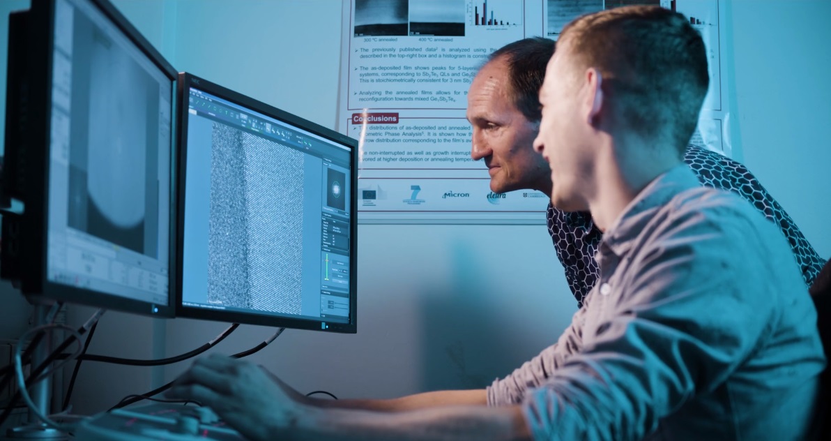
The microscope allows scientists to see the atoms in thin samples. Atomic resolution as such is not new, but this microscope is the first to combine several unique capabilities. It is able to produce images of light atoms, such as oxygen or even hydrogen, next to heavy atoms like metals. ‘We can actually see columns of hydrogen atoms in our samples,’ says Kooi. Furthermore, the microscope can distinguish between atoms of similar atomic weight. These can be identified by analysing the X rays coming from the sample, using two very large X-ray detectors.
A final unique capability of the new TEM is that it is not necessary to use a high-energy electron beam to get a good resolution. ‘Until now, the electron beam would be accelerated at 200 to 300 kilovolts,’ explains Kooi. ‘However, this can damage fragile samples. We can now get atomic resolution at 30 to 60 kilovolts, which allows us to study samples such as graphene or biological molecules.’
The microscope will primarily be used by ZIAM, which studies new materials, and CogniGron, a research institute that develops new materials for novel computer technology and approaches for a novel type of brain-inspired computers. Both institutes contributed to the price tag of the TEM of several million euros. The purchase also includes a second system: a scanning electron microscope, combined with a focused ion beam, which allows scientists to study the general structure of materials (using an electron beam) and cut out interesting sections using the ion beam, for detailed study in the new TEM.
Graphene
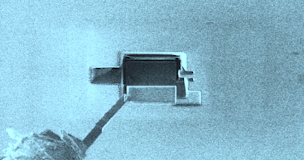
Both research institutes construct new materials, the properties of which depend on the proper alignment of atoms, for example 2D materials such as graphene, which are only one atom thick, or stacks of such 2D materials. New materials for storage memory usually have domains in the nanometre range. ‘These materials are often grown layer by layer via deposition techniques,’ explains Kooi. ‘This TEM will allow scientists to see if they properly control the atomic structures and thereby properties of their materials.’
As some of these materials are made up of oxides, being able to see the relatively light oxygen atoms is vital. The same goes for materials that could be used for the storage of hydrogen. Furthermore, the new TEM makes it possible to get a good overview and then zoom in on the atomic details. This new machine gives University of Groningen scientists access to the best possible visualization.
On 19 September, the inauguration of the ZIAM electron microscopy center will take place at the UG Zernike Institute for Advanced Materials, Nijenborgh 4, 9747 AG Groningen, with a one-day symposium, ‘The power of aberration-corrected transmission electron microscopy in materials science’. More information on the programme and registration can be found on the ZIAM website
More news
-
16 July 2026
Veni-grants for 21 UG researchers
-
-
09 July 2026
How do we keep medicines out of the environment?