Skip to ContentSkip to Navigation
Research GBB Molecular Cell Biology Research

Peroxisomal protein export

VIDI fellowship awarded to Dr. Chris Williams

Due to their vital role in cellular metabolism, peroxisomes need to adapt their protein content to changes in metabolic needs. For this reason, the ability to selectively export proteins, for degradation or targeting to other cellular compartments, can be invaluable. In contrast to other cell organelles, only a few examples of peroxisomal protein export are known and the details behind how and why proteins are exported out of peroxisomes remain largely unexplored ( Williams 2014 ).

Figure 1. Peroxisomal protein transport pathways. Schematic depicting the different transport events for both peroxisomal membrane (red) and matrix (blue) proteins. Known and putative functions of the export pathway are shown. For further reading, see (Williams 2014).

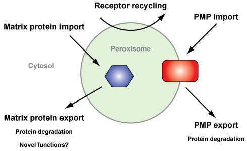
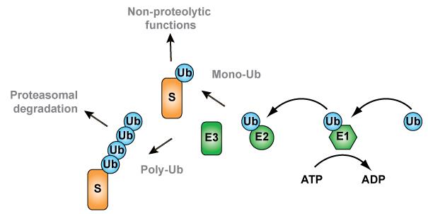
Research in the export group (Dr. Chris Williams, Chen Xin, Natasha Danda and Srishti Devarajan) focusses on the ubiquitin-dependent export and degradation of peroxisomal membrane proteins (PMPs). Our recent work demonstrates that the PMP Pex3p is specifically exported from peroxisomes and degraded upon changes to growth conditions, a process that is regulated by ubiquitination of Pex3p ( Williams and van der Klei 2013 ). Ubiquitination (Figure 2), the attachment of the protein ubiquitin to a substrate, often targets proteins for proteasome-mediated ( Tanaka 2013 ). Furthermore, Pex3p ubiquitination requires Pex2p and Pex10p; two ubiquitin ligases already implicated in peroxisomal ubiquitination events ( Williams et al, 2008 ; Platta et al, 2009 ).

Figure 2. The ubiquitination cascade. Attachment of the 8 kDa protein ubiquitin (Ub) to a substrate protein (S) is an ATP dependent process, requiring three distinct enzymes. First, the ubiquitin activating enzyme (E1) activates ubiquitin. Next, the activated ubiquitin is transferred to the active site cysteine residue of an ubiquitin conjugating enzyme (E2). Finally, an ubiquitin ligase (E3) allows conjugation of ubiquitin to the substrate. The attachment of less than four ubiquitins, often referred to as mono-ubiquitination, is usually for non-proteolytic functions. However, the ubiquitin attached to the substrate can itself become a substrate for ubiquitination, resulting in the formation of ubiquitin chains (referred to as poly-ubiquitination). Such a Ub chain can target substrates for proteasomal degradation. For further reading, see (Streich and Lima, 2014).
Our research focusses on the following:
  • By employing yeast genetics, together with various biochemical approaches, we aim to determine additional proteins involved in Pex3p ubiquitination.
  • Identify other substrates of the PMP export pathway, using a combination of proteomics and state-of-the-art fluorescence microscopy techniques.
  • Investigating the impact of PMP export on cell function, through the use of physiological approaches, combined with advanced electron microscopy/tomography and confocal laser scanning microscopy.
  • With purified proteins, we follow ubiquitination reactions in vitro using biochemical and more recently, single molecule techniques, in order to investigate the molecular mechanisms underlying PMP ubiquitination.
Last modified:19 January 2016 1.43 p.m.