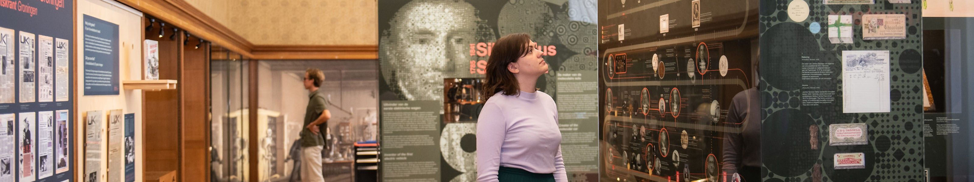
Anatomical Theatre
In the museum's anatomical theatre around 50 anatomical preparations are on display, as well as some skeletons and wax preparations. Some of the anatomical preparations are from the eighteenth century collections of Pieter de Riemer and professor Petrus Camper. Parts of the human body were prepared for educational purposes, such as a heart, a kidney or a section of bone to show the bone structure.
Anatomical Theatre
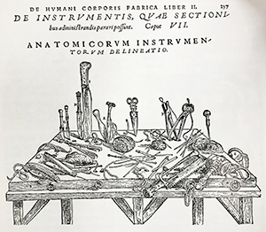
In 1615 the first anatomical lesson took place in Groningen in simple surroundings. In later years an anatomical theatre with a round gallery was built in the sacristy of the old Church of Friars Minor. An anatomical theatre was a crown jewel for a university in the seventeenth century. The museum's anatomy room is arranged like an anatomical theatre.
In an anatomical theatre, the body was put on a central dissecting table, while professors, students and commoners watched. It wasn’t just human bodies that were examined. In 1620 Nicolaas Mulerius publicly dissected a horse and a monkey.
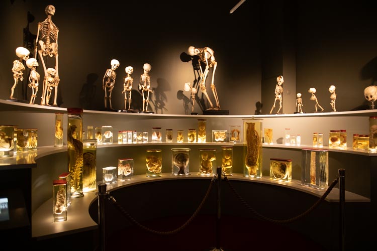
Anatomical preparations
Acquiring bodies was difficult in those days. Doctors depended on condemned criminals and strangers. Sometimes bodies were brought in from the hospice in Amsterdam, but that was only possible in winter, when conditions for preservation were good.
Because of this, a lot of time and energy was devoted to preserve body parts in alcohol. Anatomists like Pieter de Riemer and Petrus Camper acquired extensive collections of foetuses, dissected hearts and other organs preserved in alcohol. Abnormal specimens were meticulously collected too, such as Siamese twins or children suffering from hydrocephaly.
Special objects
This anatomical theatre presents some of the collection of the anatomists and doctors in Groningen. The ‘dissected man’ is especially interesting. He was a young, 27-year-old man, dissected into seven pieces around 1920. Anatomist Jan-Willem Wijhe wrote about his specimen: ‘To be able to saw through the torso like this, it is necessary to first freeze him in a mixture of ice and kitchen salt, until he’s hard like a rock; he then sounds like a bell when stricken with a hammer’
Another interesting sample is the skeleton in the middle: the man with the stooped back. He suffered from Pott’s disease, a form of tuberculosis that not only affects the lungs, but also the intervertebral discs. This results in the familiar hump.
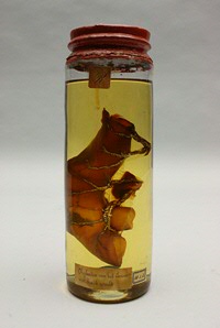
The final piece of interest is De Riemer’s human intestine specimen. The lymphatic vessels have been made visible by injecting them with mercury.
| Last modified: | 18 April 2024 3.23 p.m. |