Advent calendar - December 5th - Ceri Richards
In the Zernike Institute Advent Calendar, we are presenting 24 short spotlights in December. In these specials, we highlight PhD students, postdocs, support staff and technicians of our research groups and team - providing a glimpse in their typical day at work. In Episode 5 meet Ceri Richards, PhD student in the Molecular Biophysics group of Prof. Wouter Roos.
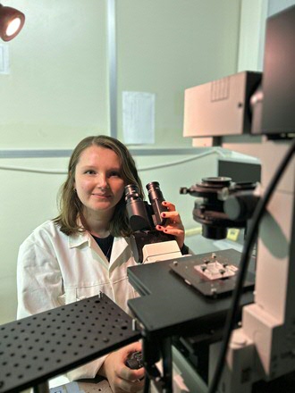
For my PhD project I work in the Molecular Biophysics group (ZIAM) with Wouter Roos and the Pharmaceutical Analysis group (GRIP) with Christoffer Åberg. Through this collaboration, we use biophysical approaches to study nanoparticles as delivery vehicles for medical applications. Nano delivery vehicles have been a hot topic for some time already, with several anti-cancer therapies using them, and more recently they were used for the Pfizer/BioNTech and Moderna corona vaccines. The idea is that the drug (or mRNA for the corona vaccines) is packaged inside of a nanoparticle which then transports it through your body and into your cells. One of the reasons why nanoparticles are such exciting candidates for drug delivery is that they come in all sorts of sizes, shapes, and materials. This makes them very versatile and tailorable. However, we do not yet understand which of the many different types of nanoparticle delivery vehicles will be most successful.
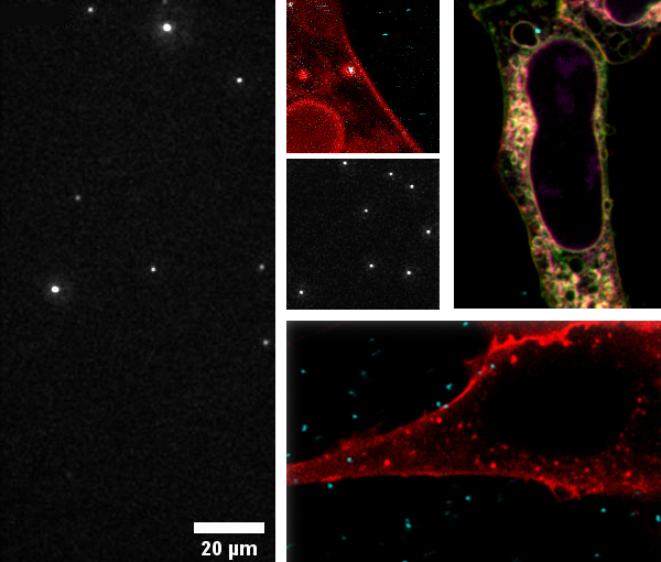
In my research, we use various fluorescence microscopy techniques to quite literally shine a light on this problem. To begin with, I used our total internal reflection fluorescence (TIRF) microscope to watch the nanoparticles adsorb to a model cell membrane called a supported lipid bilayer. This resembles the cell membrane in its fundamental components, but it is much easier to work with. We investigated the particle-membrane interactions in a methodical and strategic manner for different sized nanoparticles. After understanding this system, we used several other fluorescence-based techniques (confocal, STED and flow cytometry) to study nanoparticles interacting with living human cells.
Throughout most of my PhD I have had to balance working in two labs in two different parts of the city, which means a lot of biking around! But, on top of the exercise, it’s really nice to have the opportunity to work with researchers from different backgrounds. I also really enjoy the fact that I have access to different techniques and microscopes from the two labs. Even though fluorescence microscopy often means spending your day in a dark room, it’s worth it to see the beautiful images and videos I have captured. It is really exciting to watch the dynamic behaviour that occurs at the interface between biology and nanotechnology. With fluorescence microscopy, seeing really is believing!
| Last modified: | 01 December 2023 11.27 a.m. |
More news
-
29 April 2024
Tactile sensors
Every two weeks, UG Makers puts the spotlight on a researcher who has created something tangible, ranging from homemade measuring equipment for academic research to small or larger products that can change our daily lives. That is how UG...
-
16 April 2024
UG signs Barcelona Declaration on Open Research Information
In a significant stride toward advancing responsible research assessment and open science, the University of Groningen has officially signed the Barcelona Declaration on Open Research Information.
-
02 April 2024
Flying on wood dust
Every two weeks, UG Makers puts the spotlight on a researcher who has created something tangible, ranging from homemade measuring equipment for academic research to small or larger products that can change our daily lives. That is how UG...