Phase contrast microscope
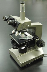
Phase contrast microscopy is an optical microscopy technique that converts phase shifts in light passing through a transparent specimen to brightness changes in the image. Phase shifts themselves are invisible, but become visible when shown as brightness variations. When light waves travels through a medium other than vacuum, interaction with the medium causes the wave amplitude and phase to change in a manner dependent on properties of the medium. Changes in amplitude (brightness) arise from the scattering and absorption of light, which is often wavelength dependent and may give rise to colors. Photographic equipment and the human eye are only sensitive to amplitude variations. Without special arrangements, phase changes are therefore invisible. Yet, phase changes often carry important information.
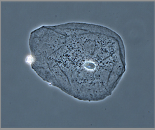
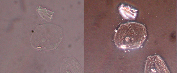
Particularly important in biology
Phase contrast microscopy is particularly important in biology. It reveals many cellular structures that are not visible with a simpler bright field microscope, as exemplified in Figure 1. These structures were made visible to earlier microscopists by staining This required additional preparation which killed the cells. The phase contrast microscope made it possible for biologists to study living cells and how they proliferate through cell division.
More information:
-
"How I discovered phase contrast" PDF Nobel Prize speech